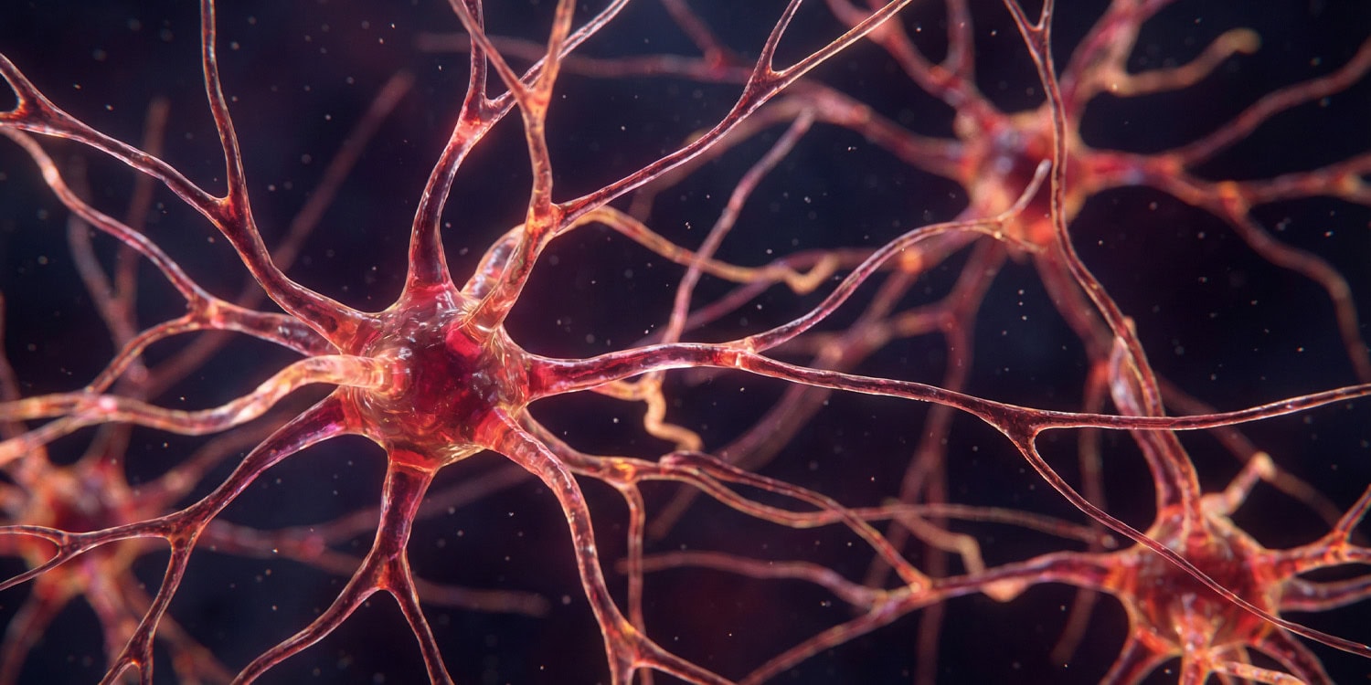
Research published in the Journal of Translational Medicine reveals distinct differences in neural communication patterns between healthy individuals and patients with Long COVID and Myalgic Encephalomyelitis/Chronic Fatigue Syndrome (ME/CFS) when experiencing cognitive load. Led by Maira Inderyas of Griffith University, the investigation specifically contrasted how the brain adapts its connectivity to maintain performance under mental exhaustion.
Healthy control participants responded to fatigue induced by a demanding Stroop task by strengthening functional connectivity, particularly between deep brain structures and the cerebellum, suggesting resource recruitment to sustain effort. Conversely, Long COVID patients displayed weakened connectivity between the nucleus accumbens, central to motivation, and the cerebellum, potentially explaining reported apathy.
ME/CFS patients exhibited separate patterns of dysfunction, including heightened connectivity within the brainstem areas governing autonomic functions, aligning with prevalent autonomic nervous system symptoms associated with that chronic illness. The team employed a powerful 7 Tesla MRI scanner to capture these subtle, high-resolution changes in functional connectivity across two testing sessions.
Researchers posit that the failure of patient groups to form the robust, integrated networks seen in healthy subjects directly contributes to cognitive impairment, commonly termed 'brain fog,' characterized by concentration and memory difficulties. This provides biological validation for subjective symptoms often previously unverified by objective testing.
In the ME/CFS cohort, the duration of the illness correlated with specific connectivity deterioration, showing a progressive weakening between the hippocampus and cerebellum over time. This suggests that the neural disruption may worsen with the chronicity of the condition, though causality remains undetermined.
While the study offers critical insight, the small sample size, common with advanced 7T imaging technology, necessitates replication for broader validation of these findings. Furthermore, the potential for prior, undiagnosed COVID-19 infections in the ME/CFS group adds a layer of complexity to the direct comparison between the two post-viral syndromes.
Ultimately, identifying these specific circuit dysfunctions through advanced imaging opens a potential avenue for developing targeted therapeutic interventions for debilitating cognitive symptoms affecting millions globally. Future longitudinal studies will aim to track whether these connectivity patterns stabilize or resolve over extended periods.